Overcome the challenges of research antibodies with next-generation reagents for target binding and detection
What are Ankyrons
Ankyrons are small (~15 kDa), single domain binding reagents based on an ankyrin-repeat scaffold. We generate Ankyron clones using a high throughput in vitro screening process, specifically selecting high affinity binders from our Teralibrary of approximately a trillion variants. These binders are recombinant by default, providing a consistent and flexible reagent to meet your research needs.
Access research areas currently hindered by a lack of reagents
Our rapidly growing Ankyron catalogue now covers more than 3000 targets with more than 16,000 recombinant clones in more than 100 species. For targets where reagents are not yet available, you can request the addition of Ankyron clones to our catalogue at no extra cost. Your target proteins (or a specific domain thereof) can be synthesized in-house as part of this process allowing for efficient and cost-effective clone generation. Please contact us to request new targets.
Emerging pathogens essential for pandemic preparedness and disease eradication
In a collaboration with the FDA, Ankyrons were rapidly generated against SARS Cov 2 spike proteins, enabling demonstration of the protective efficacy of Ankyrons against the development of viral escape mutants. This has stemmed further collaborations including provision of poliovirus, Ebola and Mpox specific Ankyron binders to allow organisation such as the CDC and Galveston National Lab at utmb health. This enables these organisations to gain key mechanistic insights into infection dynamics, driving progress toward improved systems for disease control and mitigation.
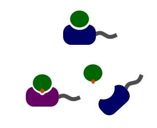
Ankyrons can be used in all the same research applications as antibodies
[table “170” not found /]
Ankyron Features
High affinity
Can be used alongside antibodies
Close proximity labeling
Cost effective and high throughput generation
Defined monoclonal sequences
Animal free
Custom generation including target protein expression available
Flexible labeling and formatting
Our catalogue single-domain Ankyrons come with a 6His-tag and a V5 epitope tag by default which can be labelled with our anti-V5 secondary reagents. V5 tags can be switched for a range of alternative labels including direct fluorescent tags and biotin, depending on your application and research goal.
Ankyron Applications
IMMUNOFLUORESCENT STAINING
Ankyrons provide excellent quality staining for immunofluorescence. Their small size means the fluorophore is in close proximity to the binding site enabling high resolution imaging. Furthermore, their small size allows for less aggressive permeabilization of cells and shorter staining and washing steps. Compared to immunofluorescence staining protocols with antibodies, the standard Ankyron protocol is significantly faster saving you over an hour in lab time. View our Ankyron protocols here. here.
Reach extracellular, intracellular and nuclear targets in immunofluorescence
[table “171” not found /]
Figure: From left to right- Huvec cells stained with clone specific for ICAM2, a cell surface molecule that are ligands for leukocyte adhesion protein LFA-1. HCT116 cells stained with clone specific for Clathrin Heavy chain 1, a key component of the clathrin protein complex involved in intracellular trafficking and spindle assembly in mitotic cells. HeLa cells stained with clone AE40370 specific for histone H3.1, a nucleosome marker
Cut down on blocking steps and save protocol time when staining with Ankyrons
Collaboration highlight: Mosquito NFKB pathway
Figure: Clone BT40163 specific to mosquito REL1A, a component of the NF-κB pathway in Ae. aegypti, and an important part of the anti-viral response.
Data kindly provided by Dr. Kevin Maringer, Pirbright Institute.
Selected Publication: Anti-Gabarap 1 Ankyron for P. berghei research
Figure: Immunofluorescent analysis with Gabarap-specific Ankyron (Clone AH50324).
HeLa cells were infected with Plasmodium berghei and the parasite was visualized with an antibody that recognises a protein in the parasitophorous vacuole membrane (PVM) in which the parasite resides in the host cell (UIS4 in magenta).
Row 1 demonstrates staining of wild type HeLa cells with the Gabarap 1 specific Ankyron. Row 2 shows the same staining but in Gabarap knock-out HeLa cells.
The Gabarap-specific Ankyron signal is in green.
Gabaraps localize to the PVM previously determined from overexpression experiments using Gabarap-GFP.
Data was kindly provided by Dr. Jacqueline Schmuckli and Prof. Volker Heussler, Institute of Cell Biology, University of Bern, SwitzerlandJ Schmuckli-Maurer, et al. Communications Biology (2024), No. 7, Art. No. 1554,“Plasmodium berghei liver stage parasites exploit host GABARAP proteins for TFEB activation”
FLOW CYTOMETRY
Quantify and assess hard-to-reach targets in flow cytometry. With their small molecular weight, Ankyrons have the advantage of efficient cellular penetration for detection of intracellular and nuclear targets without the need for harsh permeabilization.
Figure. Flow cytometric analysis of HCT116, HeLa and HUVEC following pre-conjugation of Ki67-specific Ankyron clone DS40118 with anti-V5 APC. HCT116 proliferate more rapidly than HeLa cells whereas HUVEC have a longer doubling time than either of the other cell lines, and thus lower levels of Ki67.
Figure. Ki67 in the nucleus of HeLa cells in microscopy as detected using the V5-tagged Ankyron Ki67-specific clone DS40112 (red), counterstaining nuclei with DAPI (blue) and F-actin with phalloidin (green).
Collaboration highlight: HIF-1α binders in oncology
Figure: Flow cytometry staining with HIF-1 alpha transcription factor-specific Ankyron (Clone AW40110) on NK cells from C57BL/6 mice. C57BL/6 mice were injected with MC38 tumour cells. NK cells in tumors and spleen were isolated and stained for HIF-1α. Staining with Ankyron clone AW40110 clearly shows low HIF-1α expression in the spleen which is elevated in NK cells from tumors. Data was kindly provided by Prof. Dave Withers, University of Birmingham, United Kingdom
IMMUNOHISTOCHEMISTRY
Use Ankyrons to stain fresh and FFPE tissue sections with effective tissue penetration
IHC on FFPE tissue sections using 10 μg/ml V5-tagged Ankyron and anti-V5 HRP (1 in 500), detecting with DAB (brown) and counterstaining nuclei with haematoxylin (blue-purple). Heat-mediated antigen retrieval (10 min) with Leica Tris-EDTA buffer (pH 9) using the Leica BOND RX slide staining system.
IHC Highlight: APRIL Specific Staining in Human Colon Tissue
Background APRIL (a proliferation-inducing ligand) is a member of the tumour necrosis factor (TNF) ligand superfamily (TNFSF13, also known as CD256) and plays a key role in humoral immunity, through regulating B-cell maturation, proliferation and survival by interacting with receptors TACI (TNFRSF13B) and BCMA (TNFRSF17). Under pathological conditions, APRIL is involved in the development of systemic autoimmune diseases, such as lupus erythematosus and rheumatoid arthritis. Unlike many other ligands of the TNF superfamily, APRIL can also stimulate growth of cancer cells and may be over-expressed during neoplasia. Thus, it is a useful diagnostic marker and an attractive therapeutic target in autoimmune diseases, immunoglobulin disorders and cancer.
Figure: Immunohistochemical analysis of paraffin-embedded human colon, using 10 μg/ml V5-tagged Ankyron® TNF13_HUMAN_ANK03668_3 and anti-V5 HRP (1:500), detecting with DAB (brown) and counterstaining nuclei with haematoxylin (blue-purple). Heat-mediated antigen retrieval with Leica Citrate buffer (pH6).
Watch our presentation: An introduction to the Ankyron technology
Selected Publication from the FDA: Highly specific Covid Ankyrons with functional effects
Figure: Specificity of Ankyron clones against Sars-Cov-2 spike proteins was demonstrated using Biolayer interferometry
The protective efficacy of Ankyrons against the development of viral escape mutants is comparable to that of a clinically evaluated mAb. The high-throughput generation of Ankyrons offers significant potential for rapidly developing neutralizing agents against epidemic- and pandemic-causing viruses.
Figure: SARS-CoV-2 neutralizing titres for each Ankyron were determined using the pseudovirion neutralization assay in ACE2/TMPRSS2 over expressing HEK293 cells. In this example, delta pseudovirus (1.0 X 10 6 RLU/mL) were incubated with 3-fold dilutions of variant specific Ankyrons.
Selected Animal Health Publication: The world’s first binding reagent specific to bovine IgG3
Investigating livestock immunology is important to promote sustainable farming practices, improve control of zoonotic disease and ensure food safety. The role of the IgG3 Subclass in cattle immunity was previously uncharacterised. Commercial antibodies against IgG3 were all revealed to either fail in binding or exhibit cross reactivity with other IgG subtypes. Our Ankyron technology made it possible to develop world’s first binding reagent specific for IgG3. This enabled detailed characterisation of immune response in calves administered a foot and mouse disease viral capsid vaccine.
Figure: Ankyron® AZS40101 can be used a capture or detection reagent in ELISA assays. Ankyron™ AZS40101 was used as an ELISA capture (A) or detection (B) reagent to detect recombinant bovine IgG1, IgG2 and IgG3 mAbs. Mean data ± SD for technical duplicates are shown.
A Noble, et al., Vet. Immunol. Immunother. (2024), 278, 110852,“Specificity of Ankyron clones against Sars-Cov-2 spike proteins was demonstrated using Biolayer interferometry”
Further Flexibility with the Ankyron Technology
Guided selection and p-MHC specific binders
Distinguish between targets of high homology, down to a single amino acid difference using Our guided selection strategy. During our discovery we process, we can screen two protein targets in parallel and discard the clones that bind the irrelevant protein result in selectively reactive Ankyron binders. Key examples where this selection strategy has been applied include peptide-MHC specific and idiotype specific Ankyrons.
Multimeric Ankyrons
The nature of Ankyrons as recombinat proteins with defined monoclonal sequences, expands the potential for synthesis of highly customisable binding reagents. One major example where this could be applied is in the develpment of multimeric Ankyrons, using linkers to join multiple copies of the same specificity or create multivalent binders.
As side by side comparison of Ankyrons and antibodies
Ankyrons™
Antibodies
Size (MW)
~15kD single domain
~150kD, 12 domains (IgG)
Source
Direct selection from Teralibrary™ in vitro
Immunization or selection from antibody library in vitro
Species/Isotype
Not originating from different species / no isotypes
Various species origins, various isotypes per species
Stability
Highly stable single domains
Can be highly stable for whole antibodies
Engineering
Very easy with single small domains
Possible, but more complex
Genetic definition
Monoclonal, recombinant by default
Varies, but getting from discovery to full formatted recombinant still expensive
Customization
Can be ordered as custom conjugates every time; delivered in days not months
Custom conjugation possible, but typically significant extra cost; custom reagents often have long lead times
Affinity
Typically high with direct selection from Teralibrary, bringing down the cost of high affinity reagents
Can be high if either generated as monoclonal in large animal or following selection and further optimization
Diffusion
Fast due to small 15kD domain size withe the potential for better results in IF, ICC, IHC
Limited by large 150kD size
Image resolution
Can be labeled very close to binding site, potentially offering higher resolution in microscopy
Label distance to binding site limited by size of antibody
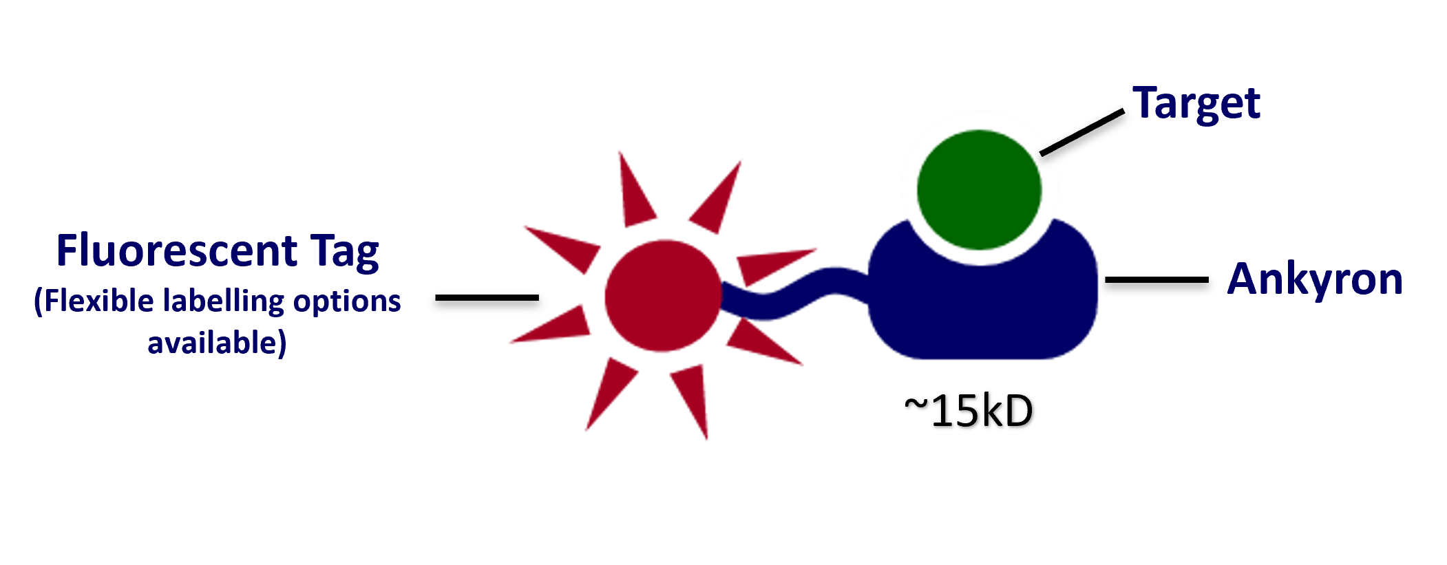


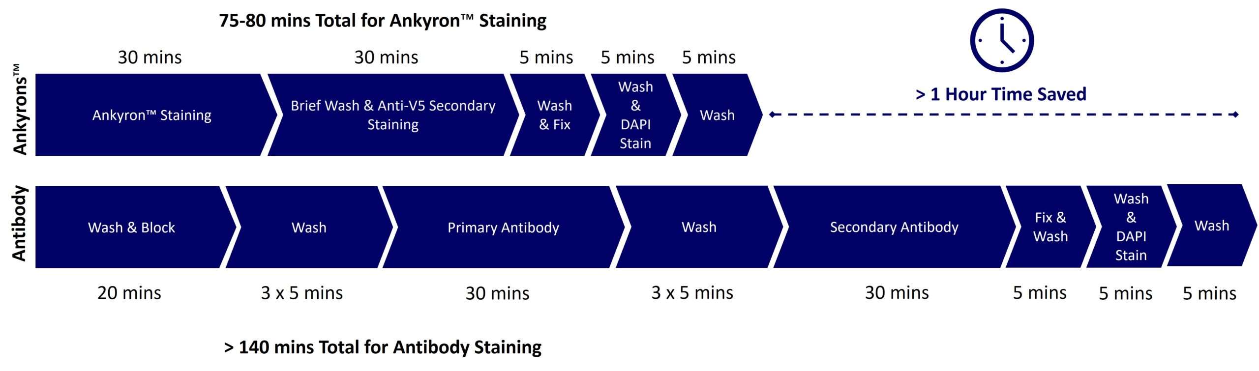

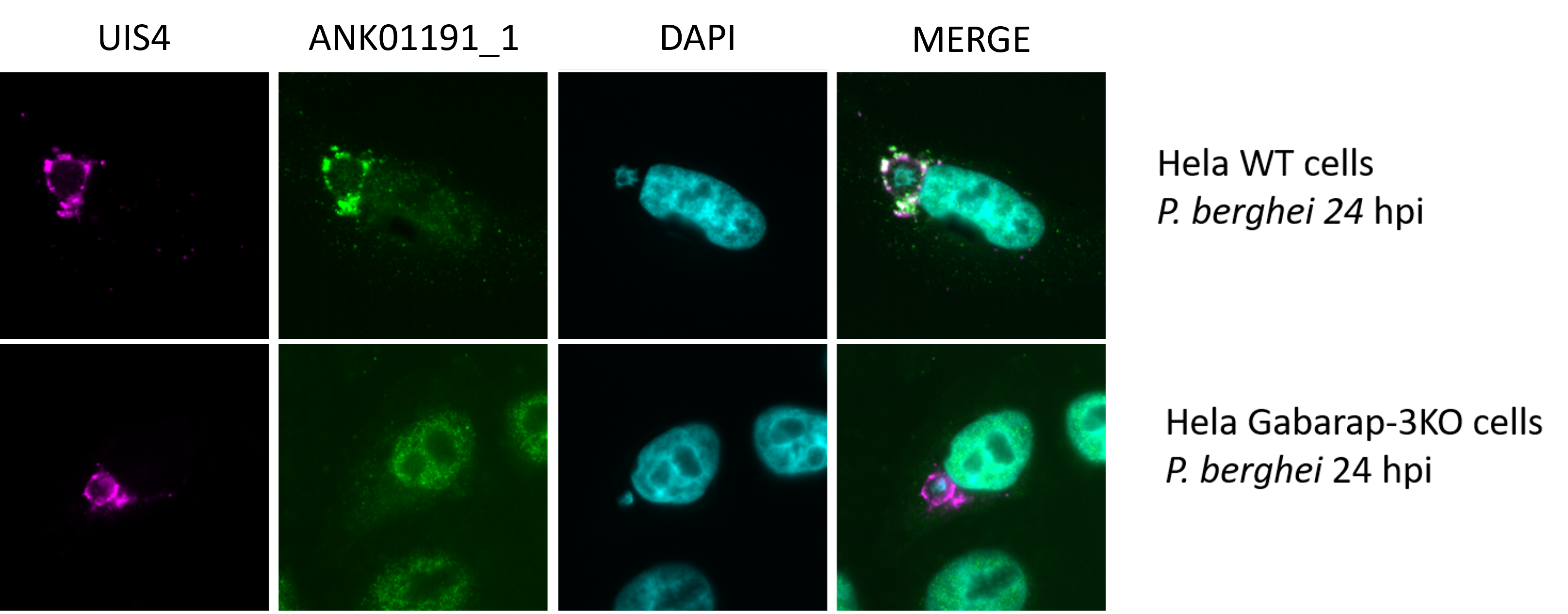 Figure: Immunofluorescent analysis with Gabarap-specific Ankyron (Clone AH50324).
HeLa cells were infected with Plasmodium berghei and the parasite was visualized with an antibody that recognises a protein in the parasitophorous vacuole membrane (PVM) in which the parasite resides in the host cell (UIS4 in magenta).
Row 1 demonstrates staining of wild type HeLa cells with the Gabarap 1 specific Ankyron. Row 2 shows the same staining but in Gabarap knock-out HeLa cells.
The Gabarap-specific Ankyron signal is in green.
Gabaraps localize to the PVM previously determined from overexpression experiments using Gabarap-GFP.
Data was kindly provided by Dr. Jacqueline Schmuckli and Prof. Volker Heussler, Institute of Cell Biology, University of Bern, Switzerland
J Schmuckli-Maurer, et al. Communications Biology (2024), No. 7, Art. No. 1554,
“Plasmodium berghei liver stage parasites exploit host GABARAP proteins for TFEB activation”
Figure: Immunofluorescent analysis with Gabarap-specific Ankyron (Clone AH50324).
HeLa cells were infected with Plasmodium berghei and the parasite was visualized with an antibody that recognises a protein in the parasitophorous vacuole membrane (PVM) in which the parasite resides in the host cell (UIS4 in magenta).
Row 1 demonstrates staining of wild type HeLa cells with the Gabarap 1 specific Ankyron. Row 2 shows the same staining but in Gabarap knock-out HeLa cells.
The Gabarap-specific Ankyron signal is in green.
Gabaraps localize to the PVM previously determined from overexpression experiments using Gabarap-GFP.
Data was kindly provided by Dr. Jacqueline Schmuckli and Prof. Volker Heussler, Institute of Cell Biology, University of Bern, Switzerland
J Schmuckli-Maurer, et al. Communications Biology (2024), No. 7, Art. No. 1554,
“Plasmodium berghei liver stage parasites exploit host GABARAP proteins for TFEB activation”

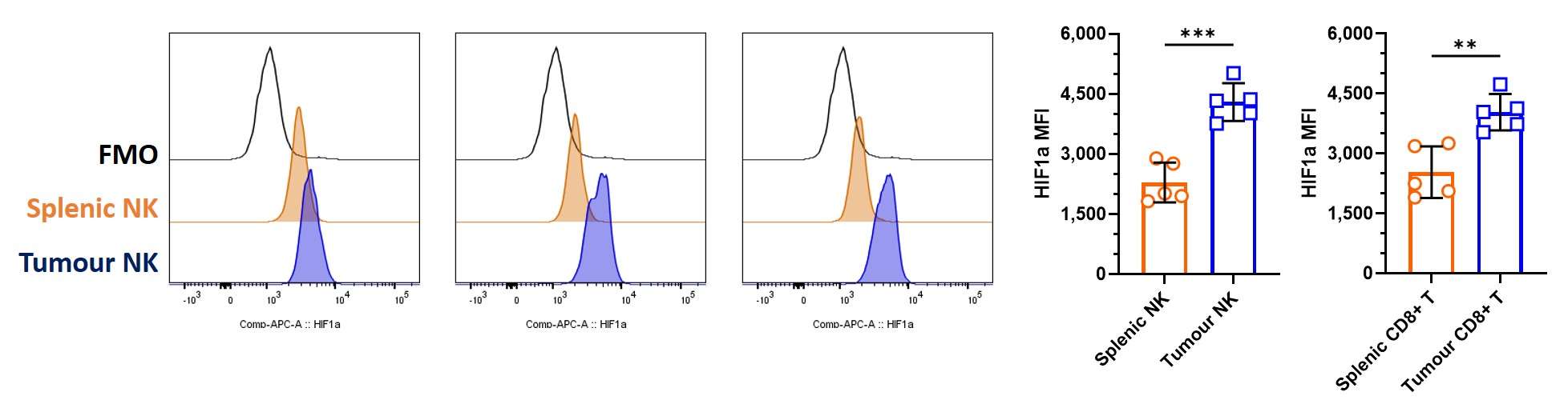


 Figure: Specificity of Ankyron clones against Sars-Cov-2 spike proteins was demonstrated using Biolayer interferometry

 Figure: Ankyron® AZS40101 can be used a capture or detection reagent in ELISA assays. Ankyron™ AZS40101 was used as an ELISA capture (A) or detection (B) reagent to detect recombinant bovine IgG1, IgG2 and IgG3 mAbs. Mean data ± SD for technical duplicates are shown.
A Noble, et al., Vet. Immunol. Immunother. (2024), 278, 110852,“Specificity of Ankyron clones against Sars-Cov-2 spike proteins was demonstrated using Biolayer interferometry”
Figure: Ankyron® AZS40101 can be used a capture or detection reagent in ELISA assays. Ankyron™ AZS40101 was used as an ELISA capture (A) or detection (B) reagent to detect recombinant bovine IgG1, IgG2 and IgG3 mAbs. Mean data ± SD for technical duplicates are shown.
A Noble, et al., Vet. Immunol. Immunother. (2024), 278, 110852,“Specificity of Ankyron clones against Sars-Cov-2 spike proteins was demonstrated using Biolayer interferometry”